Ameloblast
Ameloblasts are cells present only during tooth development that deposit tooth enamel, which is the hard outermost layer of the tooth forming the surface of the crown.
| Ameloblast | |
|---|---|
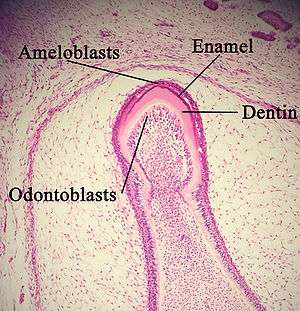 A developing tooth with ameloblasts marked. | |
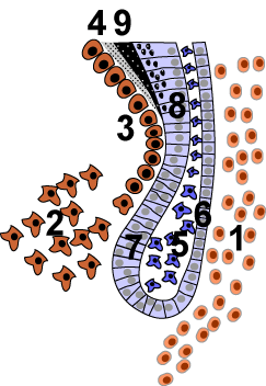 The cervical loop area: (1) dental follicle cells, (2) dental mesenchyme, (3) Odontoblasts, (4) Dentin, (5) stellate reticulum, (6) outer enamel epithelium, (7)inner enamel epithelium, (8) ameloblasts, (9) enamel. | |
| Details | |
| Identifiers | |
| Latin | ameloblastus |
| MeSH | D000565 |
| TE | E5.4.1.1.2.3.20 |
| Anatomical terms of microanatomy | |
Structure
Each ameloblast is a columnar cell approximately 4 micrometers in diameter, 40 micrometers in length and is hexagonal in cross section. The secretory end of the ameloblast ends in a six-sided pyramid-like projection known as the Tomes' process. The angulation of the Tomes' process is significant in the orientation of enamel rods, the basic unit of tooth enamel. Distal terminal bars are junctional complexes that separate the Tomes' processes from ameloblast proper.
Development
Ameloblasts are derived from oral epithelium tissue of ectodermal origin. Their differentiation from preameloblasts (whose origin is from inner enamel epithelium) is a result of signaling from the ectomesenchymal cells of the dental papilla. Initially the preameloblasts will differentiate into presecretory ameloblasts and then into secretory ameloblasts which lay down the tooth enamel. The differentiation from preameloblasts to ameloblasts occurs during the first stage of amelogenesis, called the pre-secretory (or inductive) phase. [1]
The ameloblasts will only become fully functional after the first layer of dentin (predentin) has been formed by odontoblasts. The cells are part of the reduced enamel epithelium after enamel maturation and then subsequently undergo apoptosis before or after tooth eruption.[2][3]:103 These stages occur during the third and final stage of amelogenesis, called the maturation phase.
There are various factors which can affect the differentiation and development of ameloblasts, causing abnormalities to form within the tooth structure. One example is the BMP (bone morphogenetic protein,) which has an important role in ameloblast differentiation. When Follistatin, a BMP inhibitor, is over expressed in the epithelium of developing teeth, the ameloblasts do not differentiate and no enamel forms. Another example includes the conditional deletion of 'Dicer-1' in the epithelium of developing teeth may cause impaired differentiation of ameloblasts which results in deficient enamel formation. [2]
Function
Ameloblasts are cells which secrete the enamel proteins enamelin and amelogenin which will later mineralize to form enamel, the hardest substance in the human body.[5] Ameloblasts control ionic and organic compositions of enamel. It is theorized that a circadian clock (24-hour) probably regulates enamel production on a daily cycle by the ameloblasts (similar to osteoblasts in production of bone tissue).[6] Ameloblasts adjust their secretory and resorptive activities to maintain favorable conditions for biomineralization.[3]:147
Clinical significance
These cells are sensitive to their environment. One common example is illustrated by the neonatal line, a pronounced incremental line of Retzius found in the primary teeth and in the larger cusps of the permanent first molars, showing a disruption in enamel production when the person is born.[7] High fevers in childhood are also an example of bodily stressors causing interruptions in enamel production.
Another possible example of this sensitivity (stress response pathway activation) may be the development of dental fluorosis after childhood exposure (between the ages of 2 to 8 years old) to excess consumption of fluoride, an elemental agent used to increase enamel hardness and as a result, prevent dental caries.[8]
See also
References
- He P, Zhang Y, Kim SO, Radlanski RJ, Butcher K, Schneider RA, DenBesten PK (June 2010). "Ameloblast differentiation in the human developing tooth: effects of extracellular matrices". Matrix Biology. 29 (5): 411–9. doi:10.1016/j.matbio.2010.03.001. PMC 3296366. PMID 20211728.
- Huang GT, Thesleff I (2013). Stem cells in craniofacial development and regeneration. Hoboken, New Jersey: Wiley-Blackwell. ISBN 9781118498118. OCLC 809365748.
- Nanci A (2012). Ten Cate's oral histology : development, structure, and function (Eighth ed.). Mosby. ISBN 978-0-323-24207-3.
- Takahashi S, Kawashima N, Sakamoto K, Nakata A, Kameda T, Sugiyama T, Katsube K, Suda H (February 2007). "Differentiation of an ameloblast-lineage cell line (ALC) is induced by Sonic hedgehog signaling". Biochemical and Biophysical Research Communications. 353 (2): 405–11. doi:10.1016/j.bbrc.2006.12.053. PMID 17188245.
- Gallon V, Chen L, Yang X, Moradian-Oldak J (August 2013). "Localization and quantitative co-localization of enamelin with amelogenin". Journal of Structural Biology. 183 (2): 239–49. doi:10.1016/j.jsb.2013.03.014. PMC 3737400. PMID 23563189.
- Sehic A, Nirvani M, Risnes S (October 2013). "Incremental lines in mouse molar enamel". Archives of Oral Biology. 58 (10): 1443–9. doi:10.1016/j.archoralbio.2013.06.013. PMID 23845754.
- Illustrated Dental Embryology, Histology, and Anatomy, Bath-Balogh and Fehrenbach, Elsevier, 2011, page 151
- Sierant ML, Bartlett JD (September 2012). "Stress response pathways in ameloblasts: implications for amelogenesis and dental fluorosis". Cells. 1 (3): 631–45. doi:10.3390/cells1030631. PMC 3671616. PMID 23745169.
External links
- Simmer JP, Papagerakis P, Smith CE, Fisher DC, Rountrey AN, Zheng L, Hu JC (October 2010). "Regulation of dental enamel shape and hardness". Journal of Dental Research. 89 (10): 1024–38. doi:10.1177/0022034510375829. PMC 3086535. PMID 20675598.
| Authority control |
|
|---|