Alar plate
The alar plate (or alar lamina) is a neural structure in the embryonic nervous system, part of the dorsal side of neural tube, that involves the communication of general somatic and general visceral sensory impulses. The caudal part later becomes sensory axon part of the spinal cord.
| Alar plate | |
|---|---|
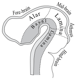 Diagram to illustrate the alar and basal laminæ of brain vesicles. | |
 aged about four and a half weeks. | |
| Details | |
| Carnegie stage | 13 |
| Precursor | Neural tube |
| Gives rise to | dorsal gray of the spinal cord, and develops into the sensory nuclei of cranial nerves V, VII, VIII, IX, and X. The inferior olivary nucleus, mesencephalic nucleus of V, and main sensory nucleus of V |
| Identifiers | |
| Latin | Lamina dorsolateralis, lamina alaris |
| TE | E5.14.1.0.1.0.4 |
| Anatomical terminology | |
The alar plate specifically later on becomes the dorsal gray of the spinal cord, and develops into the sensory nuclei of cranial nerves V, VII, VIII, IX, and X. The inferior olivary nucleus, mesencephalic nucleus of V, and main sensory nucleus of V are also developed from this plate. Also from the rhombic lip of the alar plate develops the cerebellum, which is considered to be a big exception since alar plate gives rise to sensory derivatives only.[1]
See also
References
- Siegel, Allan (2006). Essential Neuroscience. Baltimore, MD: Lippincott Williams & Wilkins. ISBN 0781750776.
This article is issued from
Wikipedia.
The text is licensed under Creative
Commons - Attribution - Sharealike.
Additional terms may apply for the media files.