Achondroplasia
Achondroplasia is a genetic disorder that results in dwarfism.[3] In those with the condition, the arms and legs are short, while the torso is typically of normal length.[3] Those affected have an average adult height of 131 centimetres (4 ft 4 in) for males and 123 centimetres (4 ft) for females.[3] Other features include an enlarged head and prominent forehead.[3] It does not affect intelligence.[3]
| Achondroplasia | |
|---|---|
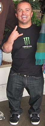 | |
| Jason "Wee Man" Acuña, an actor and stunt performer with achondroplasia | |
| Pronunciation | |
| Specialty | Medical genetics |
| Symptoms | Short arms and legs, enlarged head, prominent forehead[3] |
| Complications | Ear infections, hyperlordosis, back pain, spinal stenosis, hydrocephalus[3] |
| Causes | Mutation in FGFR3 gene[3] |
| Diagnostic method | Based on symptoms, genetic testing if uncertain[4] |
| Differential diagnosis | Hypochondroplasia, thanatophoric dysplasia, cartilage-hair hypoplasia, pseudoachondroplasia[4] |
| Treatment | Support groups, growth hormone therapy, treatment of complications[4] |
| Prognosis | 10 year shorter life expectancy[4] |
| Frequency | 1 in 27,500 people[3] |
Achondroplasia is due to a mutation in the fibroblast growth factor receptor 3 (FGFR3) gene.[3] In about 80% of cases, this occurs as a new mutation during early development.[3] In the other cases, it is inherited from one's parents in an autosomal dominant manner.[3] Those with two affected genes do not typically survive.[3] Diagnosis is generally based on symptoms but may be supported by genetic testing if uncertain.[4]
Treatments may include support groups and growth hormone therapy.[4] Efforts to treat or prevent complications such as obesity, hydrocephalus, obstructive sleep apnea, middle ear infections or spinal stenosis may be required.[4] Life expectancy of those affected is about 10 years less than average.[4] The condition affects about 1 in 27,500 people.[3] Rates are higher in Denmark and Latin America.[5] The shortest known adult with the condition is Jyoti Amge at 62.8 centimetres (2 ft 0.7 in).[6]
Signs and symptoms
- Disproportionate dwarfism
- Shortening of the proximal limbs (called rhizomelic shortening)
- Short fingers and toes with trident hands
- Large head with prominent forehead frontal bossing
- Small midface with a flattened nasal bridge
- Spinal kyphosis (convex curvature) or lordosis (concave curvature)
- Varus (bowleg) or valgus (knock knee) deformities
- Frequently have ear infections (due to Eustachian tube blockages), sleep apnea (which can be central or obstructive), and hydrocephalus
Causes
Achondroplasia is caused by a mutation in fibroblast growth factor receptor 3 (FGFR3) gene.[7] This gene is mainly responsible for making the protein, fibroblast growth factor receptor 3. This protein contributes to the production of collagen and other structural components in tissues and bones.[8] When the FGFR3 gene is mutated it interferes with how this protein interacts with growth factors leading to complications with bone production. Cartilage is not able to fully develop into bone, causing the individual to be disproportionately shorter in height.
In normal development FGFR3 has a negative regulatory effect on bone growth. In achondroplasia, the mutated form of the receptor is constitutively active and this leads to severely shortened bones. The effect is genetically dominant, with one mutant copy of the FGFR3 gene being sufficient to cause achondroplasia, while two copies of the mutant gene are invariably fatal (recessive lethal) before or shortly after birth (known as a lethal allele). A person with achondroplasia thus has a 50% chance of passing dwarfism to each of their offspring. People with achondroplasia can be born to parents that do not have the condition due to spontaneous mutation.[9]
Achondroplasia can be inherited through autosomal dominance. In couples where one partner has achondroplasia there is a 50% chance of passing the disorder onto their child every pregnancy. In situations where both parents have achondroplasia there is a 50% chance the child will have achondroplasia, 25% chance the child will not, and a 25% chance that the child will inherit the gene from both parents resulting in double dominance and leading to death.[10]
Studies have demonstrated that new gene mutations for achondroplasia are exclusively inherited from the father and occur during spermatogenesis; it has been theorized that sperm carrying the mutation in FGFR3 have a selective advantage over sperm with normal FGFR3.[11] The frequency of mutations in sperm leading to achondroplasia increase in proportion to paternal age, as well as in proportion to exposure to ionizing radiation.[12] The occurrence rate achondroplasia in the children of fathers over 50 years of age is 1 in 1875 compared to 1 in 15,000 in the general population.[13]
There are two other syndromes with a genetic basis similar to achondroplasia: hypochondroplasia and thanatophoric dysplasia.
Diagnosis

Achondroplasia can be detected before birth by prenatal ultrasound. A DNA test can be performed before birth to detect homozygosity, wherein two copies of the mutant gene are inherited, a lethal condition leading to stillbirths. Clinical features include megalocephaly, short limbs, prominent forehead, thoracolumbar kyphosis and mid-face hypoplasia.[14] Complications like dental malocclusion, hydrocephalus and repeated otitis media can be observed.[14] The risk of death in infancy is increased due to the likelihood of compression of the spinal cord with or without upper airway obstruction.
Radiologic findings
A skeletal survey is useful to confirm the diagnosis of achondroplasia. The skull is large, with a narrow foramen magnum, and relatively small skull base. The vertebral bodies are short and flattened with relatively large intervertebral disk height, and there is congenitally narrowed spinal canal. The iliac wings are small and squared, with a narrow sciatic notch and horizontal acetabular roof.[15][16] The tubular bones are short and thick with metaphyseal cupping and flaring and irregular growth plates.[15] Fibular overgrowth is present. The hand is broad with short metacarpals and phalanges, and a trident configuration. The ribs are short with cupped anterior ends.[15] If the radiographic features are not classic, a search for a different diagnosis should be entertained. Because of the extremely deformed bone structure, people with achondroplasia are often "double jointed". The diagnosis can be made by fetal ultrasound by progressive discordance between the femur length and biparietal diameter by age. The trident hand configuration can be seen if the fingers are fully extended.
Another distinct characteristic of the syndrome is thoracolumbar gibbus in infancy.
Treatment
There is no known cure for achondroplasia even though the cause of the mutation in the growth factor receptor has been found. Although used by those without achondroplasia to aid in growth, human growth hormone does not help people with achondroplasia (which involve a different hormonal pathway). However, if desired, the controversial surgery of limb-lengthening will lengthen the legs and arms of someone with achondroplasia.[17]
Usually, the best results appear within the first and second year of therapy.[18] After the second year of growth hormone therapy, beneficial bone growth decreases.[19] Therefore, GH therapy is not a satisfactory long term treatment.[18]
Complications
Children
Children with achondroplasia often have less muscle tone; because of this it is common for them to have delayed walking and motor skills. It is also common for children to have bowed legs, scoliosis, lordosis, arthritis, issues with joint flexibility, breathing problems, ear infections, and crowded teeth.[20] These issues can be treated with surgery, braces, or physical therapy.
Hydrocephalus is a severe effect associated with achondroplasia in children. This condition occurs when cerebrospinal fluid is not able to flow in and out of the skull because of how the spine narrows.[21] This fluid build up is associated with an enlarged head, vomiting, lethargy, headaches, and irritability.[22] A shunt surgery is commonly performed to treat this condition, but an endoscopic third ventriculostomy can also be done.[23]
Adults
Adults with achondroplasia often face issues with obesity and sleep apnea. It is also typical for adults to suffer from numbness or tingling in their legs because of nerve compression.
Pregnancy in women with achondroplasia is considered higher risk. Women with achondroplasia generally have their babies delivered through C-sections to prevent complications that could occur with a natural birth.[24]
Epidemiology
Achondroplasia is one of several congenital conditions with similar presentations, such as osteogenesis imperfecta, multiple epiphyseal dysplasia tarda, achondrogenesis, osteopetrosis, and thanatophoric dysplasia. This makes estimates of prevalence difficult, with changing and subjective diagnostic criteria over time. One detailed and long-running study in the Netherlands found that the prevalence determined at birth was only 1.3 per 100,000 live births.[25] Another study at the same time found a rate of 1 per 10,000.[25]
Other animals
Based on their disproportionate dwarfism, some dog breeds traditionally have been classified as "achondroplastic". This is the case for the dachshund, basset hound, corgi and bulldog breeds.[26][27][28] Data from whole genome association studies in short-limbed dogs reveal a strong association of this trait with a retro-gene coding for fibroblast growth factor 4 (FGF4).[29] Therefore, it seems unlikely that dogs and humans are achondroplastic for the same reasons. However, histological studies in some achondroplastic dog breeds have shown altered cell patterns in cartilage that are very similar to those observed in humans exhibiting achondroplasia.[30]
A similar form of achondroplasia was found in a litter of piglets from a phenotypically normal Danish sow. The dwarfism was inherited dominant in the offspring from this litter. The piglets were born phenotypically normal, but became more and more symptomatic as they reached maturity.[31] This involved a mutation of the protein Collagen, type X, alpha 1, encoded by the COL10A1 gene. In humans a similar mutation (G595E) has been associated with Schmid metaphyseal chondrodysplasia (SMCD), a relatively mild skeletal disorder that is also associated with dwarfism.
The now-extinct Ancon sheep was created by humans through the selective breeding of common domestic sheep with achondroplasia. The average-sized torso combined with the relatively smaller legs produced by achondroplasia was valued for making affected sheep less likely to escape without affecting the amount of wool or meat each sheep produced.[32]
Research
Tentative evidence as of 2019 has found that vosoritide increases growth velocity.[33]
See also
References
- "Achondroplasia". Oxford Dictionaries. Oxford University Press. Retrieved 2016-01-20.
- "Achondroplasia". Merriam-Webster Dictionary.
- "Achondroplasia". Genetics Home Reference. May 2012. Retrieved 12 December 2017.
- Pauli, RM; Adam, MP; Ardinger, HH; Pagon, RA; Wallace, SE; Bean, LJH; Mefford, HC; Stephens, K; Amemiya, A; Ledbetter, N (2012). "Achondroplasia". GeneReviews. PMID 20301331.
- "Achondroplasia". Genetic and Rare Diseases Information Center (GARD) – an NCATS Program. 2016. Retrieved 12 December 2017.
- "New world's shortest woman: It's official – Jyoti Amge from India is new record holder". Guinness World Records. 13 December 2011. Retrieved 12 December 2017.
- "Learning About Achondroplasia". National Human Genome Research Institute (NHGRI). Retrieved 2018-09-26.
- Reference, Genetics Home. "FGFR3 gene". Genetics Home Reference. Retrieved 2018-09-26.
- Richette P, Bardin T, Stheneur C (2007). "Achondroplasia: From genotype to phenotype". Joint Bone Spine. 75 (2): 125–30. doi:10.1016/j.jbspin.2007.06.007. PMID 17950653.
- "Achondroplasia". Retrieved 2018-09-26.
- Horton, William A; Hall, Judith G; Hecht, Jacqueline T (July 2007). "Achondroplasia". The Lancet. 370 (9582): 162–172. doi:10.1016/S0140-6736(07)61090-3. PMID 17630040.
- Wyrobek AJ, Eskenazi B, Young S, Arnheim N, Tiemann-Boege I, Jabs EW, Glaser RL, Pearson FS, Evenson D (2006). "Advancing age has differential effects on DNA damage, chromatin integrity, gene mutations, and aneuploidies in sperm". Proceedings of the National Academy of Sciences of the United States of America. 103 (25): 9601–9606. doi:10.1073/pnas.0506468103. PMC 1480453. PMID 16766665.
- Kovac JR, Addai J, Smith RP, Coward RM, Lamb DJ, Lipshultz LI (2013). "The effects of advanced paternal age on fertility" (PDF). Asian Journal of Andrology. 15 (6): 723–728. doi:10.1038/aja.2013.92. PMC 3854059. PMID 23912310.
- Beattie, R.M.; Champion, M.P., eds. (2004). Essential questions in paediatrics for MRCPCH (1st ed.). Knutsford, Cheshire: PasTest. ISBN 978-1-901198-99-7.
- EL-Sobky, TA; Shawky, RM; Sakr, HM; Elsayed, SM; Elsayed, NS; Ragheb, SG; Gamal, R (15 November 2017). "A systematized approach to radiographic assessment of commonly seen genetic bone diseases in children: A pictorial review". J Musculoskelet Surg Res. 1 (2): 25. doi:10.4103/jmsr.jmsr_28_17.
- "Achondroplasia Pelvis". Archived from the original on 2007-10-22. Retrieved 2007-11-28.
- Kitoh H, Kitakoji T, Tsuchiya H, Katoh M, Ishiguro N (2007). "Distraction osteogenesis of the lower extremity in patients that have achondroplasia/hypochondroplasia treated with transplantation of culture-expanded bone marrow cells and platelet-rich plasma". J Pediatr Orthop. 27 (6): 629–34. doi:10.1097/BPO.0b013e318093f523. PMID 17717461.
- Vajo, Z; Francomano, CA; Wilkin, DJ (2000). "The molecular and genetic basis of fibroblast growth factor receptor 3 disorders: the achondroplasia family of skeletal dysplasias, Muenke craniosynostosis, and Crouzon syndrome with acanthosis nigricans". Endocrine Reviews. 21 (1): 23–39. doi:10.1210/er.21.1.23. PMID 10696568.
- Aviezer, D; Golembo, M; Yayon, A (2003). "Fibroblast growth factor receptor-3 as a therapeutic target for Achondroplasia—genetic short limbed dwarfism". Current Drug Targets. 4 (5): 353–65. doi:10.2174/1389450033490993. PMID 12816345.
- "Dwarfism". kidshealth.org. Retrieved 2018-09-26.
- "Achondroplasia | Genetic and Rare Diseases Information Center (GARD) – an NCATS Program". rarediseases.info.nih.gov. Retrieved 2018-09-26.
- Kieffer, Sara. "Achondroplasia | Johns Hopkins Pediatric Neurosurgery". Retrieved 2018-09-26.
- "Hydrocephalus - Diagnosis and treatment - Mayo Clinic". www.mayoclinic.org. Retrieved 2018-09-26.
- Services, Department of Health & Human. "Dwarfism". Retrieved 2018-09-26.
- Online Mendelian Inheritance in Man (OMIM) ACHONDROPLASIA; ACH -100800
- "WebMD".
- Jones, T.C.; Hunt, R.D. (1979). "The musculoskeletal system". In Jones, T.C.; Hunt, R.D.; Smith, H.A. (eds.). Veterinary Pathology (5th ed.). Philadelphia: Lea & Febiger. pp. 1175–6. ISBN 978-0812107890.
- Willis M.B. (1989). "Inheritance of specific skeletal and structural defects". In Willis M.B. (ed.). Genetics of the Dog. Great Britain: Howell Book House. pp. 119–120. ISBN 978-0876055519.
- Parker HG, VonHoldt BM, Quignon P, et al. (August 2009). "An expressed fgf4 retrogene is associated with breed-defining chondrodysplasia in domestic dogs". Science. 325 (5943): 995–8. Bibcode:2009Sci...325..995P. doi:10.1126/science.1173275. PMC 2748762. PMID 19608863.
- Braund KG, Ghosh P, Taylor TK, Larsen LH (September 1975). "Morphological studies of the canine intervertebral disc. The assignment of the beagle to the achondroplastic classification". Res. Vet. Sci. 19 (2): 167–72. doi:10.1016/S0034-5288(18)33527-6. PMID 1166121.
- Nielsen VH, Bendixen C, Arnbjerg J, et al. (December 2000). "Abnormal growth plate function in pigs carrying a dominant mutation in type X collagen". Mamm. Genome. 11 (12): 1087–92. doi:10.1007/s003350010212. PMID 11130976.
- Gidney, Louisa (May–June 1019). "Earliest Archaeological Evidence of the Ancon Mutation in Sheep from Leicester, UK". International Journal of Osteoarchaeology. 15 (27): 318–321. doi:10.1002/oa.872. ISSN 1099-1212.
- Savarirayan, Ravi (July 4, 2019). "C-Type Natriuretic Peptide Analogue Therapy in Children with Achondroplasia". New England Journal of Medicine. 381 (1): 25–35. doi:10.1056/NEJMoa1813446. PMID 31269546.
External links
| Classification | |
|---|---|
| External resources |
| Look up achondroplasia in Wiktionary, the free dictionary. |
- Achondroplasia at Curlie
- Pauli RM (1998). "Achondroplasia". In Pagon RA, Bird TD, Dolan CR, et al. (eds.). GeneReviews. Seattle WA: University of Washington, Seattle. PMID 20301331. NBK1152.