Abrasion (dental)
Abrasion is the non-carious, mechanical wears of tooth from interaction with objects other than tooth-tooth contact.[1] It most commonly affects the premolars and canines, usually along the cervical margins.[2] Based on clinical surveys, studies have shown that abrasion is the commonest but not the sole aetiological factor for development of non-carious cervical lesions (NCCL) and is most frequently caused by incorrect toothbrushing technique.[3]
| Abrasion (dental) | |
|---|---|
 | |
| Dental abrasion | |
| Specialty | Dentistry |
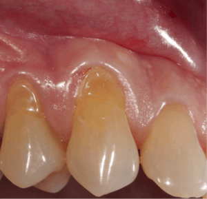
Abrasion frequently presents at the cemento-enamel junction and can be caused by many contributing factors, all with the ability to affect the tooth surface in varying degrees.[4]
The appearance may vary depending on the cause of abrasion, however most commonly presents in a V-shaped caused by excessive lateral pressure whilst tooth-brushing. The surface is shiny rather than carious, and sometimes the ridge is deep enough to see the pulp chamber within the tooth itself.
With the presence of non-carious cervical loss due to abrasion, this may lead to consequences and symptoms such as increased tooth sensitivity to hot and cold, increased plaque trapping which will result in caries and periodontal disease, difficulty of dental appliances such as retainer and denture in engaging the tooth, and also it may be aesthetically unpleasant to some people.[3]
In order for successful treatment of abrasion to occur, the cause first needs to be identified and ceased, e.g. overzealous brushing. Once this has occurred subsequent treatment may involve the changes in oral hygiene or toothpaste, application of fluoride to reduce sensitivity or the placement of a restoration to aid in reducing the progression of further tooth loss.[4]
Cause
Cause of abrasion may arise from interaction of teeth with other objects such as toothbrushes, toothpicks, floss, and ill-fitting dental appliance like retainers and dentures. Apart from that, people with habits such as nail biting, chewing tobacco, lip or tongue piercing,[5] and having occupation such as joiner, are subjected to higher risks of abrasion.
Abrasion can also occur from the type of dentifrice being utilized as some have more abrasive qualities such as whitening toothpastes. The aetiology of dental abrasion can be due to a single stimuli or, as in most cases, multi-factorial.[6] The most common cause of dental abrasion, is the combination of mechanical and chemical wear.
Tooth brushing is the most common cause of dental abrasion, which is found to develop along the gingival margin, due to vigorous brushing in this area.[7][8] The type of toothbrush, the technique used and the force applied when brushing can influence the occurrence and severity of resulting abrasion.[9] Further, brushing for extended periods of time (exceeding 2-3 min) in some cases, when combined with medium/hard bristled toothbrushes can cause abrasive lesions.[10]
Different toothbrush types are more inclined to cause abrasion, such as those with medium or hard bristles. The bristles combined with forceful brushing techniques applied can roughen the tooth surface and cause abrasion as well as aggravating the gums.[11] Repetitive irritation to the gingival margin can eventually cause recession of the gums. When the gums recede, the root surface is exposed which is more susceptible to abrasion.[12] Comparatively, electric toothbrushes have less abrasive tendencies.[13]
Types of toothpastes can also damage enamel and dentine due to the abrasive properties.[14] Specific ingredients are used in toothpaste to target removal of the bio-film and extrinsic staining however in some cases can contribute to the pastes being abrasive.[15][11] Whitening toothpastes are found to be one of the most abrasive types of toothpastes, according to the RDA Scale, detailed below.[16] In-home and clinical whitening have been proven to increase the likelihood of an individual experiencing dental abrasion. It is believed that dental abrasion due to the whitening process is caused by a combination of both mechanical and chemical irritants, for example, using whitening toothpaste and at home bleaching kits together.[16] However, if an individual is regimented in their after-whitening care then they can avoid loss of dentine minerals and in turn abrasion can be avoided. (that contribute to developing abrasion).[17]
Another factor that can contribute to abrasion is alteration of pH levels in the saliva. This can be sugary/ acidic foods and liquids. The reasoning behind this is that an increase in acidity of saliva can induce demineralization and therefore compromising the tooth structure to abrasive factors such as tooth brushing or normal wear from mastication.[18] When the tooth structure is compromised, this is where the mineral content of the saliva can create shallow depressions in the enamel and thus, when brushed can cause irreparable damage on tooth surface.[16][19][20] The dental abrasion process can be further stimulated and accelerated through the effects of dental Acid erosion.
Relative dentin abrasivity
Relative dentin abrasivity (RDA) is a standardised measurement of the abrasive effect that the components of the toothpaste have on a tooth.[10]
The RDA scale was developed by the American Dental Association (ADA). The RDA scale compares toothpaste abrasivity to standard abrasive materials and measures the depth of cut at an average of 1 millimetre per 100,000 brush strokes onto dentine.[21] This comparison generates abrasive values for the dentifrices that would be safe for daily use.[12] In vitro dental studies showed a positive correlation between the highest RDAs and greater dentin wear.[22]
Since 1998, the RDA value is set by the standards DIN EN ISO 11609. Currently, the claim on products such as toothpaste are not regulated by law, however a dentifrice is required to have a level lower than 250 to be considered safe and before being given the ADA seal of approval.[23] The values obtained depend on the size, quantity and surface structure of abrasive used in toothpastes.
While the RDA score has been shown to have a statistically significant correlation to the presence of abrasion, it is not the only contributing factor to consider.[21][12][24] Other factors such as the amount of pressure used whilst brushing, the type, thickness and dispersion of bristle in the toothbrush and the time spent brushing are other factors that contribute to dental abrasion.[24]
Treatment
There are several reasons to treat abrasion lesion(s) (also known as ‘Class V cavity’) such as:-
- Sensitivity.
- Presence of carious lesion.
- Aesthetically unpleasant.
- Arresting the progression of the lesion.
- Reducing potential onset of caries or periodontal disease as these lesions can present as a plaque retention factor.
- Where there is a risk of pulpal exposure if lesion depth is severe enough.
- When retention of a removable appliance is interfered. Ie. Denture
- To improve denture clasp(s) retention.
- Overall integrity of tooth structure is compromised.
In order for successful treatment of abrasion to occur, the aetiology first needs to be identified. The most accurate way of doing so is completing a thorough medical, dental, social and diet history. All aspects needs to be investigated as in many cases the cause of abrasion can be multi-factorial. Once a definitive diagnosis is completed the appropriate treatment can commence. Treatment for abrasion can present in varying difficulties depending on the current degree or progress caused by the abrasion. Abrasion often presents in conjunction with other dental conditions such as attrition, decay and erosion. Evidence suggest there is a decrease in the effect of dental abrasion with dental erosion when fluoride varnish is applied onto teeth.[25] Successful treatment focuses on the prevention and progression on the condition and modifies the current habit/s instigating the condition.
Removal of Causes
If the cause of abrasion is due to habitual behaviours, the discontinuation and change of habit is critical in the prevention of further tooth loss.[26] The correct brushing technique is pivotal and involves a gentle scrub technique with small horizontal movements with an extra-soft/soft bristle brush.[24] Excessive lateral force can be corrected by holding the toothbrush in a pen grasp or by using the non-dominant hand to brush.[24] If abrasion is the result of an ill-fitting dental appliance, this should be corrected or replaced by a dental practitioner and should not be attempted in a home setting.
Chemical
The current selection of dentifrice should also be critically analysed and changed to include a less abrasive and gentler paste such as sensitive toothpaste as evidence suggests that a very abrasive toothpaste would lead to loss of tooth structure.[27] A toothpaste containing increased fluoride will also help combat the increased sensitivity and risk to dental decay.[28] Fluoride varnish is known to alleviate hypersensitivity in teeth and can be used as a preventive measure for high risk patients of dental erosion with abrasion because fluoride varnish is reported to have an effect on the surface and subsurface of the tooth.[29]
Treatment in the dental chair may include a fluoride application or the placement of a restoration in more severe cases. If the lesion is small and confined to enamel or cementum, a restoration is not warranted, instead the eradication of rough edges should occur to reduce plaque retentive properties.[30] However, in the case of dental decay, aesthetic concerns or defects close to the pulp a restoration may be completed.[31] Further restorative work may be required when the lesion compromises the overall strength of the tooth or when the defect contributes to a periodontal problem the lesion may be restored.[32]
Once abrasions has been diagnosed and treated it should be closely monitored to identify further progression or potential relief of symptoms.
Restoration
Ideal properties of restoration materials particularly for these lesions include:-[33]
- Satisfactory wear resistance most commonly caused by overzealous/excessive force used during toothbrushing.
- Low modulus of elasticity, given that teeth (anterior dentition) have been considered to flex around the cervical area (area closest to gum levels).
- Good aesthetics.
There are other properties of restoration materials which could be considered appropriate, although not specific to Class V restorations, which includes:-
- Small filler particles for polishability to achieve better aesthetics.
- Sufficiently stiff consistency to hold shape but still allows easy handling for placement into a cavity.
- Self-curing/setting or curable to any depth.
- Dimensionally stable or low shrinkage/stress.
- Fluoride release.
- Self-adhesive to enamel and dentine.
Dental materials such as amalgam, glass ionomer (GI), resin-modified glass ionomer (a variant of GI) and resin composite are the types of restoration materials available when active treatment by means of restoration is appropriate.
Taking into consideration these factors and their respective dental materials' properties, evidence and studies has shown that resin-modified glass ionomer (RMGI) restoration material is the recommended restoration material in clinical situations as it performs optimally - provided when aesthetics is not the top priority when restoring these lesions.[33] The surface of such lesions should be roughened prior to its restoration[34][35][36][37][38] - whether material is GI-based or resin-based[33] - with no need for bevelling of the coronal aspect of the cavity.[35][39][40]
See also
References
- López-Frías, Francisco J.; Castellanos-Cosano, Lizett; Martín-González, Jenifer; Llamas-Carreras, José M.; Segura-Egea, Juan J. (2012-02-01). "Clinical measurement of tooth wear: Tooth wear indices". Journal of Clinical and Experimental Dentistry. 4 (1): e48–e53. doi:10.4317/jced.50592. ISSN 1989-5488. PMC 3908810. PMID 24558525.
- Forbes-Haley, C., Jones, S. B., Davies, M., & West, N. X. (2016). Establishing the Effect of Brushing and a Day's Diet on Tooth Tissue Loss in Vitro. Dentistry Journal, 4 (3).
- Perez, Cesar dos Reis; Gonzalez, Mariana Rodrigues; Prado, Natália Aráujo Silva; Miranda, Marianna Sorozini Ferreira de; Macêdo, Mariana de Andrade; Fernandes, Bárbara Monteiro Pessôa (2012). "Restoration of Noncarious Cervical Lesions: When, Why, and How". International Journal of Dentistry. 2012: 687058. doi:10.1155/2012/687058. ISSN 1687-8728. PMC 3246729. PMID 22216032.
- Sugita L, Nakashima S, Ikeda A, Burrow M, Nikaido T. A pilot study to assess the morphology and progression of non-carious cervical lesions. Journal of Dentistry 2017:51-6.
- De Moor, R J G; Witte, A M J C De; Delmé, K I M; Bruyne, M A A De; Hommez, G M G; Goyvaerts, D (October 2005). "Dental and oral complications of lip and tongue piercings". British Dental Journal. 199 (8): 506–509. doi:10.1038/sj.bdj.4812852. ISSN 1476-5373. PMID 16244618.
- Lee A, He LH, Lyons K, Swain MV. Tooth wear and wear investigations in dentistry. Journal of oral rehabilitation. 2012 Mar 1;39(3):217-25.
- Sadaf D, Ahmad Z. Role of brushing and occlusal forces in non-carious cervical lesions (NCCL). International journal of biomedical science: IJBS. 2014 Dec;10(4):265.
- Salas MM, Nascimento GG, Vargas-Ferreira F, Tarquinio SB, Huysmans MC, Demarco FF. Diet influenced tooth erosion prevalence in children and adolescents: Results of a meta-analysis and meta-regression. Journal of dentistry. 2015 Aug 31;43(8):865-75.
- Zanatta FB, Bergoli AD, Werle SB, Antoniazzi RP. Biofilm removal and gingival abrasion with medium and soft toothbrushes. Oral Health Prev Dent. 2011 Jan 1;9(2):177-83.
- Vieira GH, Nogueira MB, Gaio EJ, Rosing CK, Santiago SL, Rego RO. Effect of Whitening Toothpastes on Dentin Abrasion: An In Vitro Study. Oral health & preventive dentistry. 2016 Jun 27.
- Wiegand A, Kuhn M, Sener B, Roos M, Attin T. Abrasion of eroded dentin caused by toothpaste slurries of different abrasivity and toothbrushes of different filament diameter. Journal of dentistry. 2009 Jun 30;37(6):480-4.
- Addy M, Hunter ML. Can tooth brushing damage your health? Effects on oral and dental tissues. International dental journal. 2003 Jun 1;53(S3):177-86.
- Wiegand A, Lemmrich F, Attin T. Influence of rotating–oscillating, sonic and ultrasonic action of power toothbrushes on abrasion of sound and eroded dentine. Journal of periodontal research. 2006 Jun 1;41(3):221-7.
- Tellefsen G, Liljeborg A, Johannsen A, Johannsen G. The role of the toothbrush in the abrasion process. International journal of dental hygiene. 2011 Nov 1;9(4):284-90.
- Bizhang M, Riemer K, Arnold WH, Domin J, Zimmer S. Influence of Bristle Stiffness of Manual Toothbrushes on Eroded and Sound Human Dentin–An In Vitro Study. PLoS ONE. 2016 Apr 12;11(4):e0153250.
- Demarco FF, Meireles SS, Sarmento HR, Dantas RV, Botero T, Tarquinio SB. Erosion and abrasion on dental structures undergoing at-home bleaching. Clin Cosmet Investig Dent. 2011 Jul 18;3:45-52.
- John SS, White DJ. History of the development of abrasivity limits for dentifrices 2015
- Neel EA, Aljabo A, Strange A, Ibrahim S, Coathup M, Young AM, Bozec L, Mudera V. Demineralization–remineralization dynamics in teeth and bone. International Journal of Nanomedicine. 2016;11:4743.
- Lussi A, Schlüter N, Rakhmatullina E, Ganss C. Dental erosion–an overview with emphasis on chemical and histopathological aspects. Caries research. 2011 May 31;45(Suppl. 1):2-12.
- Jaeggi T, Lussi A. Toothbrush abrasion of erosively altered enamel after intraoral exposure to saliva: an in situ study. Caries Research. 1999 Sep 23;33(6):455-61.
- John S, White D. History of the development of abrasivity limits for dentifrices. The journal of clinical dentistry. 2015;26(02):51-54.
- Macdonald E, North A, Maggio B, Sufi F, Mason S, Moore C, Addy M, West NX. Clinical study investigating abrasive effects of three toothpastes and water in an in situ model. J Dent 2010;38:509–516.
- Toothpastes. Ada.org. 2017 [cited 7 May 2017]. Available from: http://www.ada.org/en/member-center/oral-health-topics/toothpastes
- Walsh M, Darby ML. Dental hygiene: theory and practice. Elsevier Health Sciences; 2014 Apr 15.
- Sar Sancakli, H.; Austin, R. S.; Al-Saqabi, F.; Moazzez, R.; Bartlett, D. (March 2015). "The influence of varnish and high fluoride on erosion and abrasion in a laboratory investigation". Australian Dental Journal. 60 (1): 38–42. doi:10.1111/adj.12271. ISSN 1834-7819. PMID 25721276.
- Bergström J, Lavstedt S. An epidemiologic approach to toothbrushing and dental abrasion. Community dentistry and oral epidemiology. 1979 Feb 1;7(1):57-64.
- Ganss, C.; Marten, J.; Hara, A. T.; Schlueter, N. (November 2016). "Toothpastes and enamel erosion/abrasion - Impact of active ingredients and the particulate fraction". Journal of Dentistry. 54: 62–67. doi:10.1016/j.jdent.2016.09.005. ISSN 1879-176X. PMID 27650640.
- Seong J, Parkinson CP, Davies M, Claydon NC, West NX. Randomised clinical trial to evaluate changes in dentine tubule occlusion following 4 weeks use of an occluding toothpaste. Clinical Oral Investigations. 2017 Apr 1:1-9.
- Sar Sancakli, H.; Austin, R. S.; Al-Saqabi, F.; Moazzez, R.; Bartlett, D. (March 2015). "The influence of varnish and high fluoride on erosion and abrasion in a laboratory investigation". Australian Dental Journal. 60 (1): 38–42. doi:10.1111/adj.12271. ISSN 1834-7819. PMID 25721276.
- Marinho VC. Cochrane fluoride reviews: an overview of the evidence on caries prevention with fluoride treatments. Faculty Dental Journal. 2014 Apr;5(2):78-83.
- Harpenau LA, Noble WH, Kao RT. Diagnosis and management of dental wear. Today's FDA: official monthly journal of the Florida Dental Association. 2011 Dec;24(5):50-7.
- White JM, Eakle WS. Rationale and treatment approach in minimally invasive dentistry. The Journal of the American Dental Association. 2000 Jun 30;131:13S-9S.
- Burke, F. J. (2015). Dental Materials: What Goes Where? Class V Restorations. Dental Update,42(9), 829-839.
- Stewardson DA, Creanor S, Thornley P, Bigg T, Bromage C, Browne A, Cottam D, Dalby D, Gilmour J, Horton J, Roberts E, Westoby L, Burke T. The survival of class V restorations in general dental practice: part 3. five year survival. Br Dent J 2012; 212: E14.
- Heintze SD, Ruffieux C, Rousson V. Clinical performance of cervical restorations − a meta-analysis. Dent Mater 2010; 26: 993−1000.
- Van Dijken JWV. A prospective 8-year evaluation of a mild two-step self-etching adhesive and a heavily filled two-step etchand-rinse system in non-carious cervical lesions. Dent Mater 2010; 26: 940−948.
- Gwinnett AJ, Kanca J. Interfacial morphology of resin composite and shiny erosion layers. Am J Dent 1992; 5: 316−317.
- Tay FR, Pashley DH. Resin bonding to cervical sclerotic dentine: a review. J Dent 2004; 32: 173−179.
- Da Costa TEF, Loguercio AD, Reis A. Effect of enamel bevel on the clinical performance of resin composite restorations placed in noncarious cervical lesions. J Esthet Restor Dent 2013; 25: 346−356.
- Schroeder M, Reis A, Luque-Martinez I, Loguercio AD, Masterson D, Maia LC. Effect of enamel bevel on retention of cervical composite resin restorations: a systematic review and meta-analysis. J Dent 2015; 43: 777−788.