3D ultrasound
3D ultrasound is a medical ultrasound technique, often used in fetal, cardiac, trans-rectal and intra-vascular applications. 3D ultrasound refers specifically to the volume rendering of ultrasound data and is also referred to as 4D (3-spatial dimensions plus 1-time dimension) when it involves a series of 3D volumes collected over time.
When generating a 3D volume the ultrasound data can be collected in four common ways. Freehand, which involves tilting the probe and capturing a series of ultrasound images and recording the transducer orientation for each slice. Mechanically, where the internal linear probe tilt is handled by a motor inside the probe. Using an endoprobe, which generates the volume by inserting a probe and then removing the transducer in a controlled manner. The fourth technology is the matrix array transducer that uses beamsteering to sample points throughout a pyramid shaped volume.[1]
Risks
The general risks of ultrasound also apply to 3D Ultrasound. Essentially ultrasound is considered safe. While other imaging modalities use radioactive dye or ionizing radiation, for example, ultrasound transducers send pulses of high frequency sound into the body and then listen for the echo.
In summary, the primary risks associated with ultrasound would be the potential heating of tissue or cavitation. The mechanisms by which tissue heating and cavitation are measured are through the standards called thermal index (TI) and mechanical index (MI). Even though the FDA outlines very safe values for maximum TI and MI it is still recommended to avoid unnecessary ultrasound imaging.[2]
Applications
Obstetrics
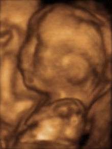
The progress of growing babies in the womb is difficult to examine as the fetuses are always in motion with rapid rate of heartbeat. So while scanning a womb, the patients need to keep their breath still so the real picture can be captured. With 3D US, the doctors can now detect the movement of fetuses in a flash of second and also take necessary step immediately. In addition to examining the position of the fetus, the problems and any abnormal behaviors in the womb can be detected like accumulation of fluid or any spine curvature.[3] With 3D US and 3D surfacing the heartbeat of the fetus can also be traced enabling the doctor to outline the growth of the baby.[4]
Cardiology
The application of three-dimensional ultrasound in cardiac treatments has done outstanding progress in scanning and treating the heart issues. When the 3D ultrasound technology is used to visualize the cardiac state of an individual, it is called as 3D Echocardiography.[5] With the integration of other technologies with the Ultrasound, it is now possible to track the quantitative measures like the chamber volume that occurs during the cardiac cycle. Also, it provides other useful information like tracking the blood flow, speed of contractions and expansions.[6] With the 3D Echocardiography method doctors now can easily detect the artery diseases and can finely examine the various defects. The echo applications helps to give a real-time image of the cardiac structures.[7]
Surgical guidance
Traditionally, with the 2D ultrasound, the specific position of organs and tissues useful in surgeries could not be located especially in the oblique plane. However, with the advent of 3D ultrasound, the imaging technique has evolved manifolds enabling the surgeons to get a real-time picture of the tissues and organs and visualize the complete scan more efficiently.[8] In addition to this, 3D ultrasound provides surgical guidance in terms of treating transplant and cancer with its technique of rotational visualizing while scanning.[9] This technology has evolved various methods like rotational scanning, slice projection, use of integrated array transducer that helps the surgeons to handle the traumatic cancer patients.[10] Further, with 3D ultrasound doctors can now treat various kind of tumors as every tissue can be easily diagnosed and introspected to study the defects and cause.[11] Thus, we see that the ultimate scanning and visualization done by 3D ultrasound has developed better ways of treating the patients with problems like cancer, tumors and transplants.
Vascular imaging
Blood vessels and arteries movements are not easy to track because of their distribution due to which capturing a real-time picture becomes difficult. Diagnosis is widely used in every kind of treatment and with the 3D US, it is now possible to track dynamic movement of blood cells, veins and arteries.[12] In addition to this, various types of diagnosis like measuring the diameter, diagnosing the wall between the arteries can be detected with a magnetic tracker integrated with 3D US that helps in accurate positioning.[13] So the technology gives imaging assistance and also has a sensor that helps in tracking the position of vessels in operations.
Regional anesthesia
Real-time three-dimensional ultrasound is used during peripheral nerve blockade procedures to identify relevant anatomy and monitor the spread of local anesthetic around the nerve. Peripheral nerve blockades prevent the transmission of pain signals from the site of injury to the brain without deep sedation, which makes them particularly useful for outpatient orthopedic procedures. Real-time 3D ultrasound allows muscles, nerves and vessels to be clearly identified while a needle or catheter is advanced under the skin. 3D ultrasound is able to view the needle regardless of the plane of the image, which is a substantial improvement over 2D ultrasound. Additionally, the image can be rotated or cropped in real time to reveal anatomical structures within a volume of tissue. Physicians at the Mayo Clinic in Jacksonville have been developing techniques using real time 3D ultrasound to guide peripheral nerve blocks for shoulder, knee, and ankle surgery.[14][15]
References
- Hoskins, Peter; Martin, Kevin; Thrush, Abigail (2010). Diagnostic ultrasound : physics and equipment (2nd ed.). Cambridge, UK: Cambridge University Press. ISBN 978-0-521-75710-2.
- Health, Center for Devices and Radiological. "Medical Imaging - Ultrasound Imaging". www.fda.gov.
- Baba, Kazunori; Okai, Takashi; Kozuma, Shiro; Taketani, Yuji (1999). "Fetal Abnormalities: Evaluation with Real-time-Processible Three-dimensional US—Preliminary Report". Radiology. 211 (2): 441–446. doi:10.1148/radiology.211.2.r99mr02441. PMID 10228526.
- Acar, Philippe; Battle, Laia; Dulac, Yves; Peyre, Marianne; Dubourdieu, Hélène; Hascoet, Sébastien; Groussolles, Marion; Vayssière, Christophe (2014). "Real-time three-dimensional foetal echocardiography using a new transabdominal xMATRIX array transducer". Archives of Cardiovascular Diseases. 107 (1): 4–9. doi:10.1016/j.acvd.2013.10.003. PMID 24364911.
- Huang, Qinghua; Zeng, Zhaozheng (2017). "A Review on Real-Time 3D Ultrasound Imaging Technology". BioMed Research International. 2017: 1–20. doi:10.1155/2017/6027029. PMC 5385255. PMID 28459067.
- Pedrosa, J.; Barbosa, D.; Almeida, N.; Bernard, O.; Bosch, J.; d'Hooge, J. (2016). "Cardiac Chamber Volumetric Assessment Using 3D Ultrasound - A Review". Current Pharmaceutical Design. 22 (1): 105–21. doi:10.2174/1381612822666151109112652. PMID 26548305.
- Picano, E.; Pellikka, P. A. (2013). "Stress echo applications beyond coronary artery disease". European Heart Journal. 35 (16): 1033–1040. doi:10.1093/eurheartj/eht350. PMID 24126880.
- Yan, P. (2016). "SU-F-T-41: 3D MTP-TRUS for Prostate Implant". Medical Physics. 43 (6Part13): 3470–3471. doi:10.1118/1.4956176.
- Ding, Mingyue; Cardinal, H. Neale; Fenster, Aaron (2003). "Automatic needle segmentation in three-dimensional ultrasound images using two orthogonal two-dimensional image projections". Medical Physics. 30 (2): 222–234. doi:10.1118/1.1538231. PMID 12607840.
- Mahboob, Syed; McPhillips, Rachael; Qiu, Zhen; Jiang, Yun; Meggs, Carl; Schiavone, Giuseppe; Button, Tim; Desmulliez, Marc; Demore, Christine; Cochran, Sandy; Eljamel, Sam (2016). "Intraoperative Ultrasound-Guided Resection of Gliomas: A Meta-Analysis and Review of the Literature" (PDF). World Neurosurgery. 92: 255–263. doi:10.1016/j.wneu.2016.05.007. PMID 27178235.
- Moiyadi, Aliasgar V.; Shetty, Prakash (2016). "Direct navigated 3D ultrasound for resection of brain tumors: A useful tool for intraoperative image guidance". Neurosurgical Focus. 40 (3): E5. doi:10.3171/2015.12.FOCUS15529. PMID 26926063.
- Jin, Chang-zhu; Nam, Kweon-Ho; Paeng, Dong-Guk (2014). "The spatio-temporal variation of rat carotid artery bifurcation by ultrasound imaging". 2014 IEEE International Ultrasonics Symposium. pp. 1900–1903. doi:10.1109/ULTSYM.2014.0472. ISBN 978-1-4799-7049-0.
- Pfister, Karin; Schierling, Wilma; Jung, Ernst Michael; Apfelbeck, Hanna; Hennersperger, Christoph; Kasprzak, Piotr M. (2016). "Standardized 2D ultrasound versus 3D/4D ultrasound and image fusion for measurement of aortic aneurysm diameter in follow-up after EVAR". Clinical Hemorheology and Microcirculation. 62 (3): 249–260. doi:10.3233/CH-152012. PMID 26484714.
- "Real-Time 3-D Ultrasound Speeds Patient Recovery" (Press release). Mayo Clinic. July 13, 2007. Retrieved May 21, 2014.
- Feinglass, Neil G.; Clendenen, Steven R.; Torp, Klaus D.; Wang, R Doris; Castello, Ramon; Greengrass, Roy A. (2007). "Real-Time Three-Dimensional Ultrasound for Continuous Popliteal Blockade: A Case Report and Image Description". Anesthesia & Analgesia. 105 (1): 272–274. doi:10.1213/01.ane.0000265439.02497.a7. PMID 17578987.
External links
- The History of Ultrasounds (including 3D ultrasounds)
- The Endowment for Human Development provides numerous 4D ultrasounds that can be viewed online.
- Scans uncover secrets of the womb BBC News
- About the discovery of medical ultrasonography
- Learn how to perform 3D/4d Ultrasounds
- Safety Concerns Details a number of health concerns over this unregulated practice
- 3D Ultrasound Clinic and Education Facility A 3D Ultrasound
clinic in Sacramento, CA that specializes in performing 3D ultrasound and training sonographers.
| X-ray/ Radiography |
| ||||||||||||
|---|---|---|---|---|---|---|---|---|---|---|---|---|---|
| MRI | |||||||||||||
| Ultrasound | |||||||||||||
| Radionuclide |
| ||||||||||||
| Optical/Laser | |||||||||||||
| Thermography |
| ||||||||||||
| Target conditions |
| ||||||||||||
| |||||||||||||